|
|
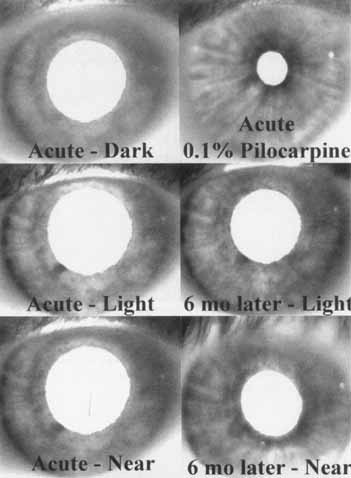
| Fig. 11 Six infrared transillumination views of the same iris in a patient with acute Adie's affecting all but one of the segments. The middle and lower photographs on the fight are from the same eye 6 months later. The photographs on the left side show that there is only one segment (the dark area at the pupil border at the 7:00 position) that still contracts appropriately to light and near. This example was chosen because the rest of the iris sphincter-reacting segment can be discerned in the dark (captured during the latency period of the pupil light reflex), in light, and at near. In the acute-dark photograph (top left), the area at the 7:00 meridian cannot be seen but becomes dark in response to light (middle left, acute light) and in response to low concentration pilocarpine (top right). All the denervated segments show darkening to the 0.1% pilocarpine except for the one segment that normally is innervated, and therefore presumably not supersensitive, which appears colored at the 7:30 meridian (top right, acute 0.1% pilocarpine). This same patient was examined 6 months later (right side, middle and bottom) and shows a light-near dissociation with a darkening on near response. The pupil is slightly smaller in light than in the acute state because of some sustained firing of accommodative fibers that have started to reinnervate the sphincter areas. (Reprinted with permission from Kardon RH, Corbett JJ, Thompson HS: Segmental denervation and reinnervation of the iris sphincter as shown by infrared videographic transillumination. Ophthalmology 105:313, 1998) |