|
|
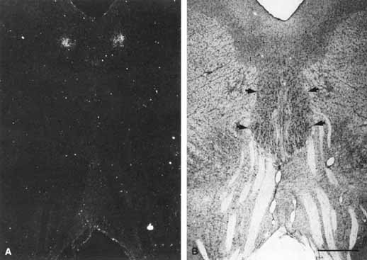
| Fig. 4 A. (Left) Transneuronal autoradiographic label in the Edinger–Westphal nuclei, seems bilaterally adjacent to the midline, ventral to the cerebral aqueduct. B. (Right) The label seen in A corresponds on each side to the fairly distinct cell group (thin arrows), the lateral visceral cell column of the Edinger–Westphal nucleus, shown in a Nissl-counterstained section. The somatic subnuclei of the oculomotor complex (thick arrows) contain larger nuclei. Fascicles from the oculomotor complex are seen streaming inferiorly toward the interpeduncular fossa. Scale bar = 1 mm. (Reprinted with permission from Kourouvan HD, Horton JC: Transneuronal retinal input to the primate Edinger–Westphal nucleus. J Comp Neurol 381:68, 1997) |