|
|
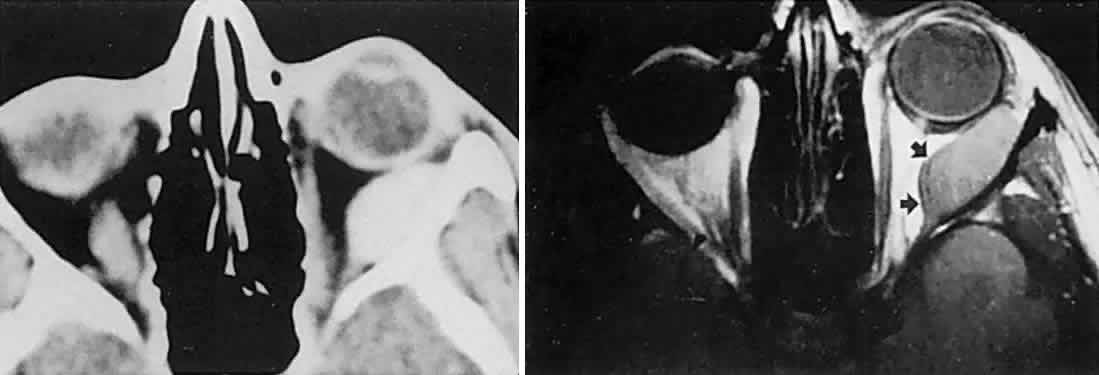
| Fig. 21. Young patient had slowly evolving unilateral proptosis. Left. Enhanced CT scan shows laterally placed homogeneous mass. Right. MRI with contact coil shows mass well separated from optic nerve and splaying lateral rectus (arrows) on medial surface of lesion; tumor was a fibrous histiocytoma. |