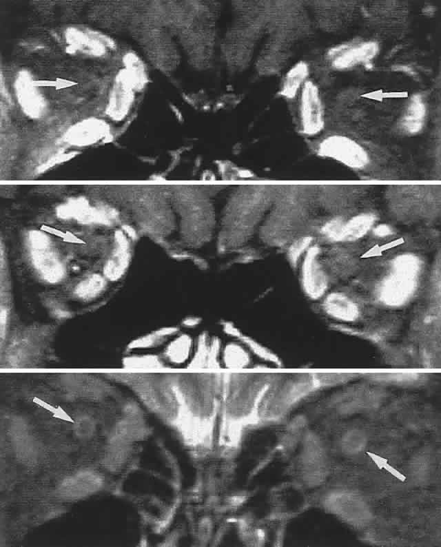
Fig. 20.
MRI of orbits, coronal sections. Fat suppression.
Top.
Midorbit.
Middle.
Orbital apex.
Bottom.
T2-weighted.
Arrows
indicate optic nerve; note ring of CSF (
bottom
).