|
|
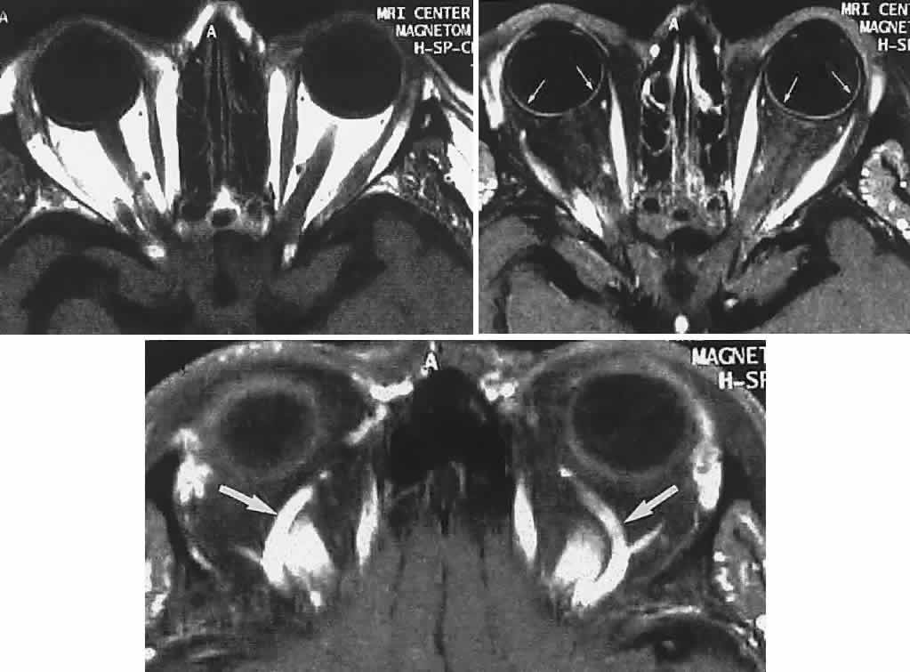
| Fig. 19. MRI of orbits, axial sections. Top. T1-weighted: orbital fat is white (hyperintense), muscles are dark. Middle. Fat saturation with gadolinium through midorbit: orbital fat signal suppressed (dark), accentuates hyperintense muscles; note also choroid (small arrows). Bottom. Fat-saturation technique through superior aspect of orbit; note superior ophthalmic veins (arrows). |