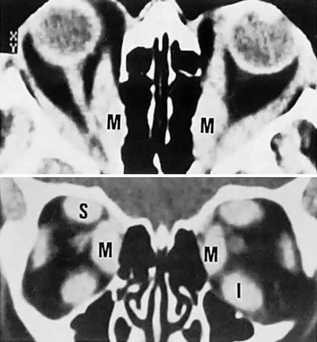
Fig. 18.
CT scan in Graves' disease.
Top.
Axial section shows massively enlarged horizontal recti (
M, medial
) with packed apex.
Bottom.
Coronal section demonstrates enlarged medial (
M
), superior (
S
), and inferior (
I
) recti.