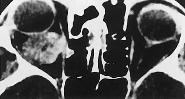
Fig. 17.
CT scan shows typical configuration of intraconal hemangioma. Note slightly inhomogeneous content, rounded distinct borders, and clear apical space.