|
|
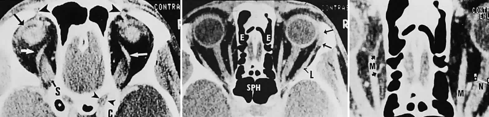
| Fig. 16. Contrast-enhanced CT scan of normal orbits. Top. Superior orbit section shows superior ophthalmic veins (white arrows), superior rectus origin (S), left levator muscle complex (black arrow), position of trochlea and tendon of superior oblique muscles (large arrowheads), right optic canal (small arrowheads), and anterior clinoid (C). (*, pneumatized left anterior clinoid.) Middle. Midorbital section shows ethmoidal sinus complex (E), sphenoidal sinus (SPH), lacrimal gland (arrows), and lateral rectus (L). Bottom. Enlargement shows left medial rectus (M), with anterior (top arrow) and posterior (bottom arrow) ethmoidal arteries; note cursor across optic nerve on right (N). |