|
|
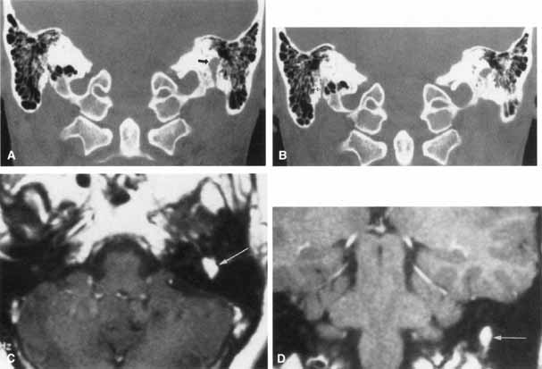
| Fig. 16 Patient with facial neuroma. A. Coronal CT scan demonstrating enlarged stylomastoid foramen and facial nerve mass (arrow). B. Note size of canal in Figure A (black arrow) compared with normal sized canal (open arrow). C. Axial MRI scan showing an enhancing mass of tympanic segment (arrow). D. Coronal gadolinium-enhanced MRI section demonstrating same mass in mastoid segment (arrow). |