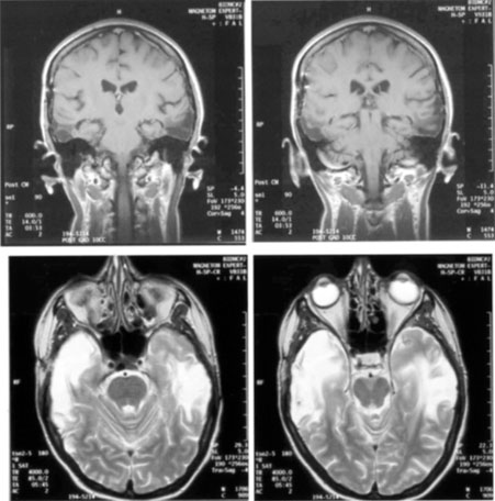
Fig. 27.
Coronal (
top
) and axial (
bottom
) MRI of a patient with associative prosopagnosia from bilateral anterior temporal lesions.