|
|
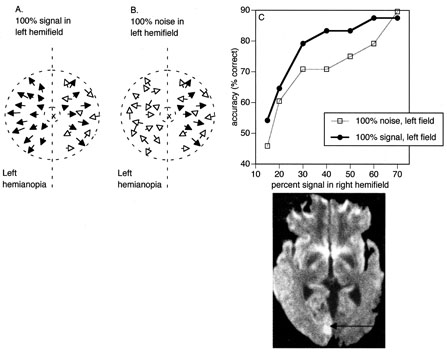
| Fig. 17. Interactions between blind and seeing hemifields. Bottom shows the diffusion weighted MRI of the small right striate lesion, causing left hemianopia. Left shows the two contrasting stimuli. The patient's task is to judge the direction of the optic flow motion (black arrows) in the seeing right hemifield, from amidst a background of moving noise (white arrows). In the blind field there is either random noise or a strong optic flow motion pattern. Results on right show that the accuracy of the patient at all levels of mixed noise and optic flow are improved by 5% to 10% when there is a strong flow pattern in the blind field. |