|
|
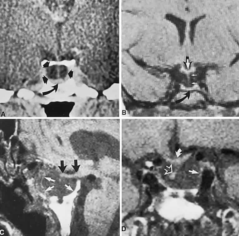
| Fig. 20. Neuroimaging of empty sella. A. Computed tomography scan of a 46-year-old woman with a vague headache complex. Coronal section shows a dark empty sella (arrows) with a preserved midline hypophyseal stalk and flattened remnant of the pituitary gland (curved arrow). B. T1-weighted magnetic resonance imaging (MRI) shows the flattened pituitary (curved arrow), the midline stalk (small white arrows), and the chiasm above (large white arrow). C. MRI (TR, 800 milliseconds; TE, 26 milliseconds); sagittal section shows a large, remodeled sella (white arrows) with moderate prolapse of the optic nerves and chiasm (black arrows). D. Coronal section through the sella; solid arrow on remaining dural wall, open arrow on distorted pituitary stalk, with the chiasm above (small curved arrow). |