|
|
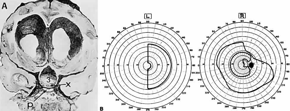
| Fig. 18. Effect of internal hydrocephalus. A. Coronal section of the brain and basal structures from a patient with a cerebellar tumor and optic atrophy. The third ventricle (3) is dilated, the chiasm (X) is stretched, and the sella with the pituitary (P) is compressed. B. Visual fields of a 17-year-old girl with postmeningitic hydrocephalus. At surgery, the ballooned third ventricle was seen to stretch the chiasm; ventriculostomy through the anterior wall of the third ventricle relieved hydrocephalus with improvement in field defects. (From ref. 208) |