|
|
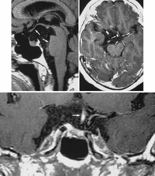
| Fig. 12. Suprasellar cyst presenting with chronic visual loss. Magnetic resonance imaging (MRI) shows that the signal is isointense to cerebrospinal fluid. Top. Left, sagittal section demonstrates elevation of the optic chiasm and posterior deformation of the midbrain. Right, axial computed tomography displays leftward displacement of the pituitary stalk (arrow). Bottom. MRI shows lateral compression of the stalk (arrow). |