|
|
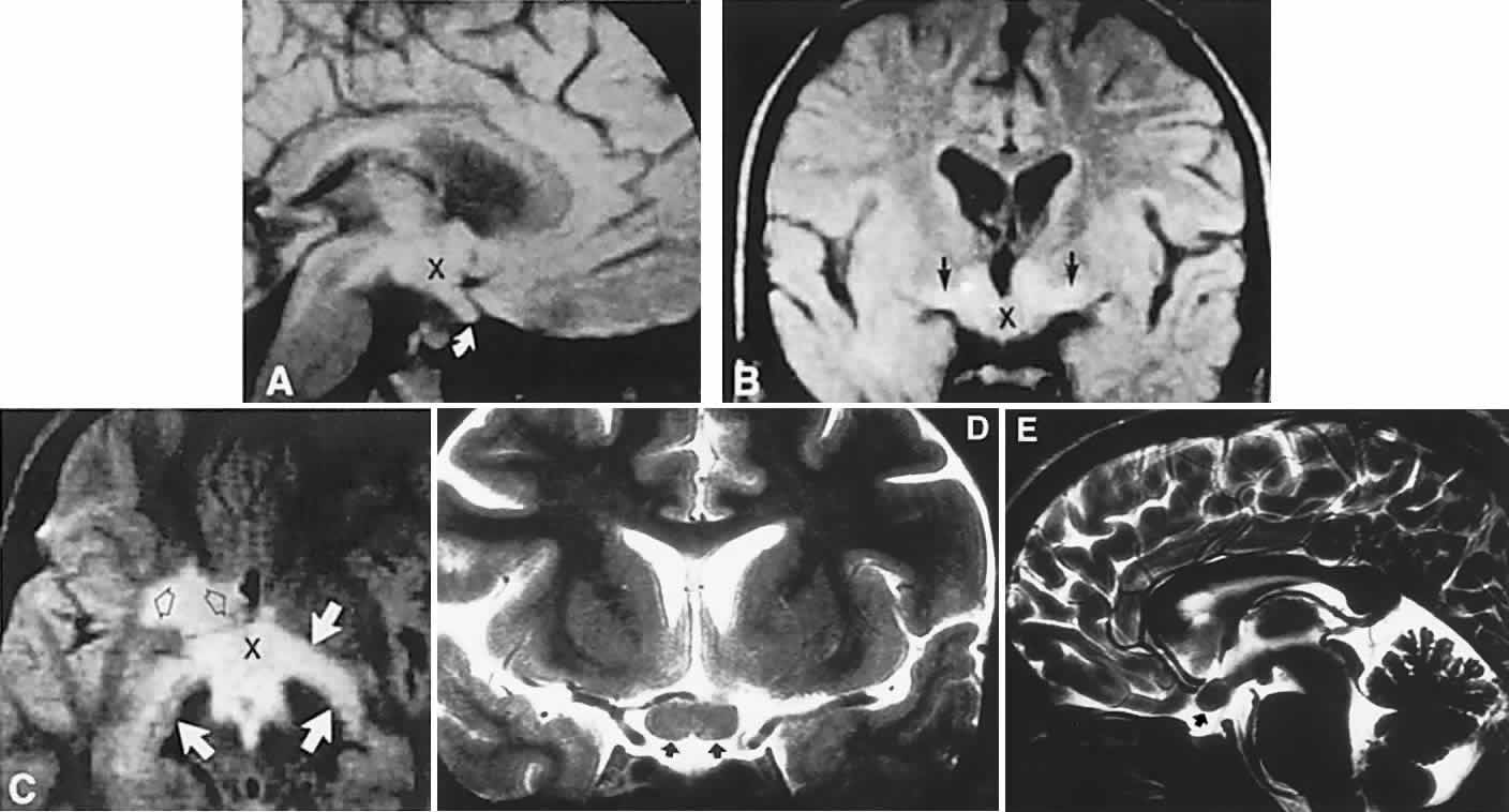
| Fig. 8. Magnetic resonance imaging of a childhood optochiasmatic glioma. A. Sagittal section shows a ballooned optic nerve (curved arrow) and a thickened chiasm (X). B. Coronal section reveals a glioma in the chiasm (X), optic tracts (arrows), and walls of the third ventricle. C. T2-weighted axial image (TR, 200 milliseconds; TE 60 milliseconds) with postbiopsy edema (open arrows) and a glioma in the chiasm (X) with posterior extension to both optic tracts (white arrows). Coronal (D) and sagittal (E) views of a glioma of the chiasm (arrows). |