|
|
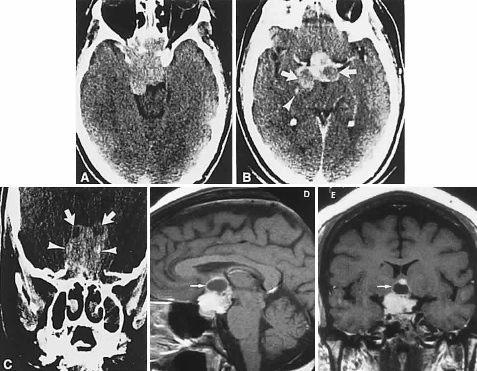
| Fig. 7. Computed tomography scan of a large, multicystic craniopharyngioma. A. Axial section through the sella shows destruction of the bony skull base. Axial (B) and coronal (C) sections show cysts (white arrows) and calcification (arrowheads). Contrast-enhanced magnetic resonance imaging of the craniopharyngioma. Sagittal (D) and coronal (E) sections with gadolinium show solid and cystic (arrows) portions. |