|
|
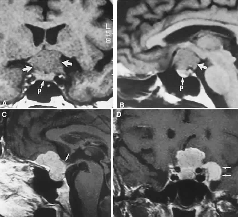
| Fig. 4. Magnetic resonance imaging of a suprasellar meningioma (TR, 600 milliseconds; TE, 20 milliseconds). A. Coronal section of a large meningioma (large arrows), isodense to brain. B. Sagittal section. Note the normal sella and pituitary gland (p). Sagittal (C) and coronal (D) sections of a planum meningioma, extending into the sella. Note the upward deflection of the chiasm (arrow in C) and extension to the cavernous sinus (arrows in D). |