|
|
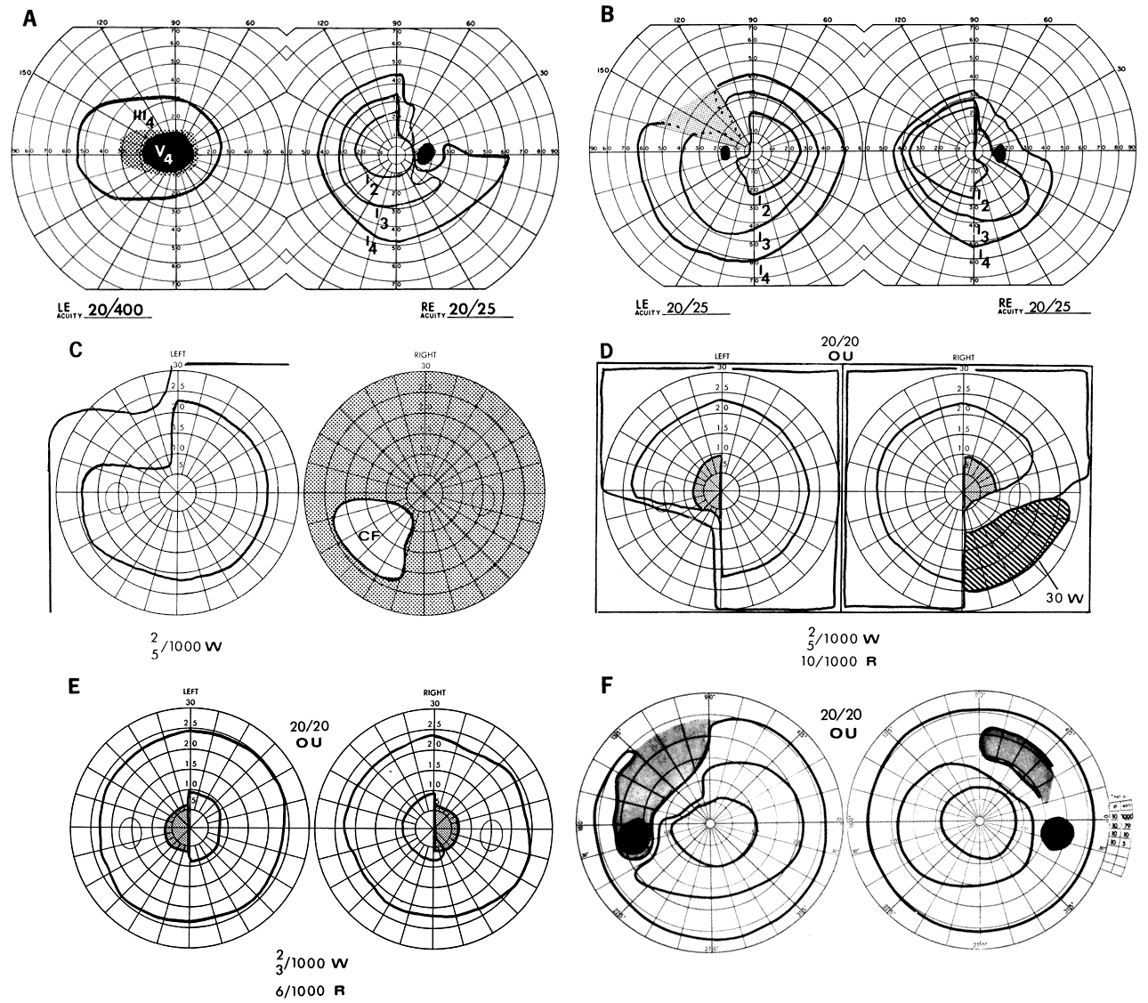
| Fig. 1. Chiasmal field defects. A. “Junctional scotoma” combines typical optic nerve defect in the left field (LE) with temporal hemianopia in the right (RE) (see also C). B. Classic bitemporal hemianopia. Riddoch's phenomenon (motion perception) is demonstrable in the shaded area of the left field. C. Asymmetric progression, with severe visual deficit in the right eye and early superior temporal depression on the left. D. Defect characteristic of posterior chiasmal notch lesion. Note the central hemianopic scotomas and inferior quadrant defects. E. Central hemianopic scotomas typical of posterior chiasmal interference. F. Temporal hemianopic arcuate scotomas. The patient sustained head trauma, with resultant field defects and diabetes insipidus. |