|
|
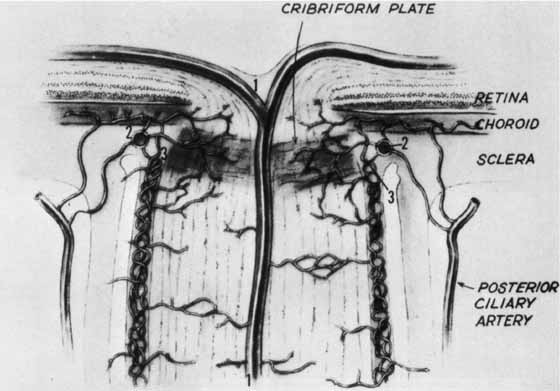
| Fig. 4 Blood supply of the optic nerve head. 1, central retinal artery; 2, arterial circle of Zinn-Haller; 3, pial arterial network. Contribution to Zinn-Haller circle from posterior ciliary arteries, pial plexus, and peripapillary choroids; the latter also sends branches directly to prelaminar disc substance. (Modified from Kolker AE, Hetherington J Jr: Becker-Shaffer's diagnosis and therapy of the glaucomas, ed 3. St. Louis: Mosby, 1970) |