|
|
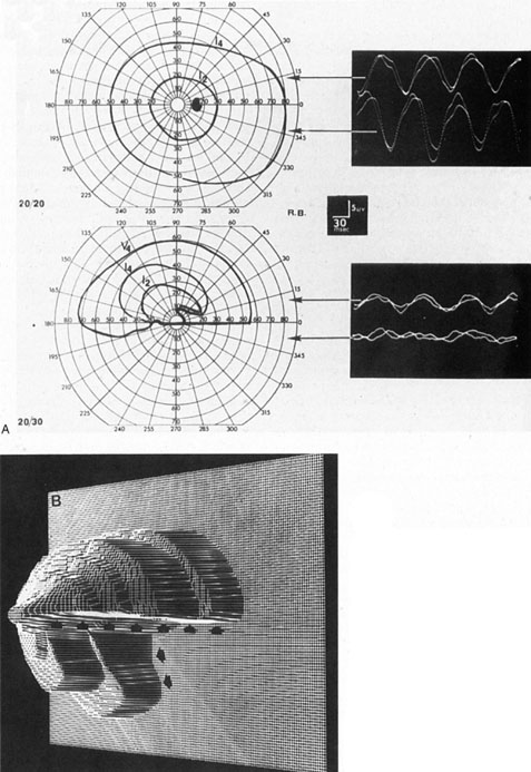
| Fig. 7. Dense inferior altitudinal field defect resulting from anterior ischemic optic neuropathy of left eye. A. Goldmann visual fields and VEP tracings for right eye (top), which is normal, and left eye (middle), which shows almost complete loss of the lower hemifield. Steady-state VEPs are in response to 8 Hz pattern reversal stimulation of upper or lower half of the visual field and are normal and symmetrical in both half-fields of the right eye and are diminished, especially in the inferior field, of the left eye. B. Three-dimensional computer reconstruction field defect in the left eye; arrows define the sharp edge of the absolute defect. (Courtesy H. Stanley Thompson, MD.) |