|
|
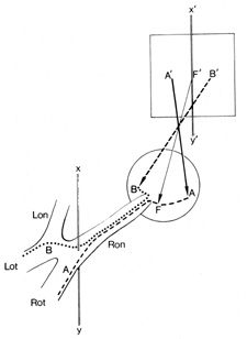
| Fig. 6. Visual field of the right eye divided into a temporal (B') and nasal (A') hemifield, by a vertical line (X', Y') through the point of fixation (F'). There is no anatomic or functional segregation of crossed (nasal retinal) fibers, B (···········), and uncrossed (temporal retinal) fibers, A (−−−−−−−), before the junction of the optic nerve with the chiasm at the vertical line (X,Y). Therefore, lesions anterior to the chiasm produce defects that extend across the vertical, whereas chiasmal and retrochiasmal lesions produce defects confined to one hemifield. Lon, left optic nerve; Lot, left optic tract; Ron, right optic nerve; Rot, right optic tract. |