|
|
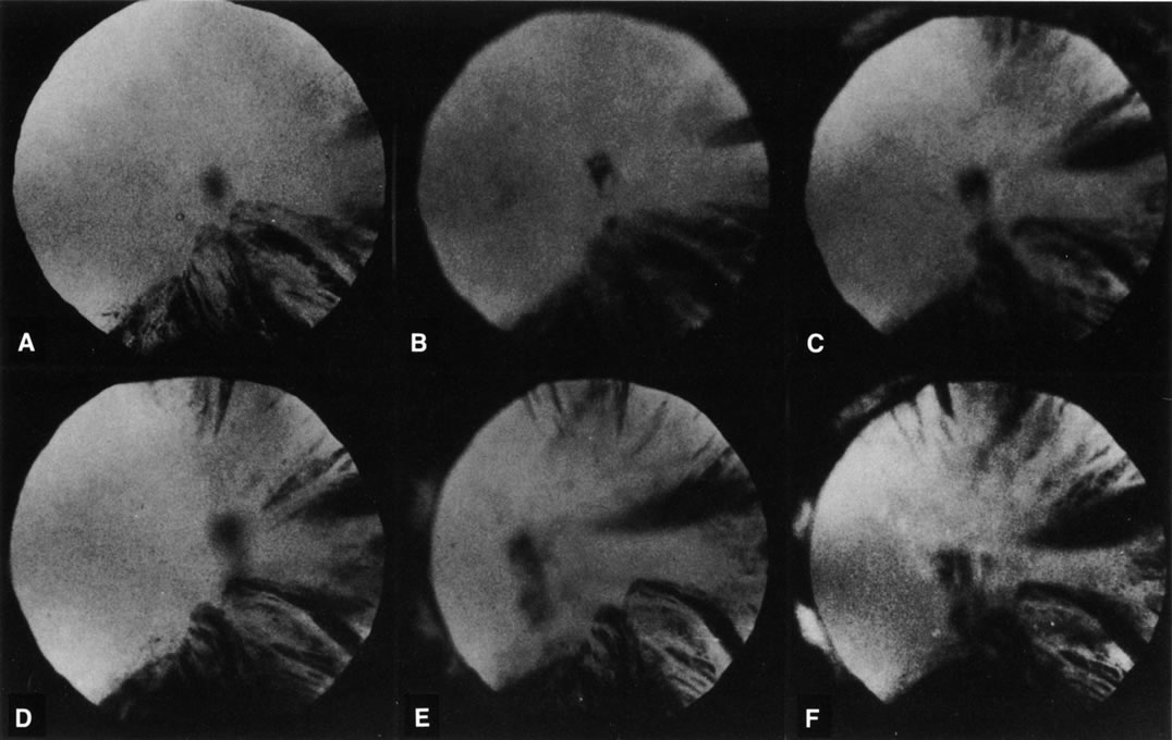
| Fig. 8. Follow-up retroillumination photographs of an eye with a cortical cataract, obtained at various intervals. A. First visit (1-21-87); B. 1 year later (1-27-88); C. 22 months later (11-14-88); D. 28 months later (12-4-89); E. 35 months later; F. 41 months later (6-18-90). With such photographs one may be able to plot the progression rate of a cortical cataract, and aid in performing longitudinal studies. Note the central opacity, which is out of focus and represents a small posterior subcapsular cataract. |