|
|
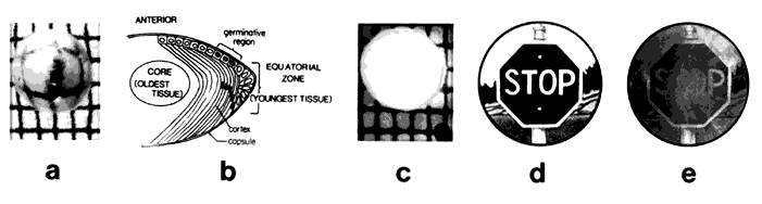
| Fig. 2. Clear and cataractous lens. a. Clear lens allows an unobstructed view of the wire grid placed behind it. b. Cartoon of the structure of the lens. The anterior surface of the lens has a unicellular layer of epithelial cells (youngest tissue). Cells at the anterior equatorial region divide and migrate to the cortex as they are overlaid by less-mature cells. These cells produce most the crystallins. As develop-ment and maturation proceed, the cells denucleate and elongate. Tissue originally found in the embry-onic lens is found in the core or nucleus (oldest tissue). c. The cataractous lens prohibits viewing the wire grid behind it. d. Artist's view through a clear uncolored young lens. The image is clear and crisp. e. Artist's view through a lens with developing cataract. The image is partially obscured,and the field is darkened due to browning of the lens that accompanies aging. (Taylor A, Dorey CK, Nowell T Jr. Oxidative stress and ascorbate in relation to risk for cataract and age-related maculopathy. In Packer L, Fuchs J [eds]: Vitamin C in Health and Disease. New York: Marcel Dekker,1997.) |