|
|
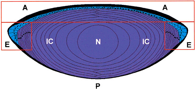
| Fig. 1. Cross-sectional view of the mammalian lens. The lens is morphologically an asymmetric structure with a single layer of epithelial cells at the anterior surface. The anterior cells in Section A, although coupled to each other, do not appear to be well coupled to the fibers beneath nor does there appear to be many junctional complexes between fiber cells in this region. Cells in the equatorial section (E) do appear to be well coupled at the fiber-fiber and fiber-epithelium interface. Functionally, therefore, the lens appears to behave as two symmetric structures superimposed. The anterior (leaky) epithelium represents one system and the fiber cells represent the other. This model accounts for the radial distribution of currents around the lens and also explains why only a small asymmetry voltage is observed in double-chamber experiments. The posterior surface (P) has no epithelial cells, and the differentiated fibers in the inner cortex (IC) and nucleus (N) have lost their organelles. See text for further details. |