|
|
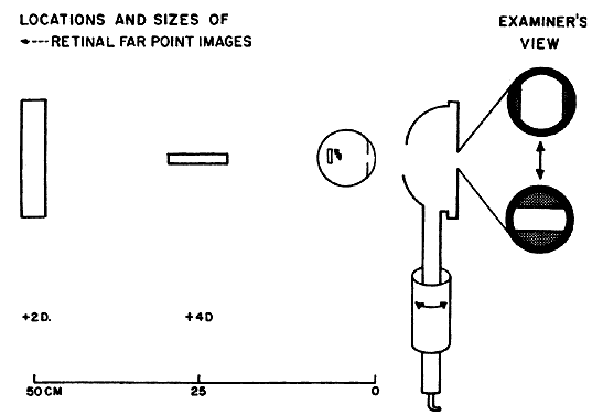
| Fig. 25. Optics of meridional comparison for astigmatism Astigmatism is detected by observing meridional differences in the sizes of virtual retinal images projected into the far-point plane of a hyperopic eye. The diagram (not to scale) shows differences in the far point image sizes for an eye with compound hyperopic astigmatism requiring a net correction of + 2.00 D, + 2.00 D × 180 degrees. The unfocused filament image of the retinoscope is shown projected onto the retina in the vertical meridian only. The retina serves as an object for the images focused in the far-point plane of each meridian. Note that the largest image results from magnification because of its being focused at twice the distance from the nodal point of the eye as the smaller image. The scale measures distance (cm) posterior to the cornea. On the right are shown the retinal images as viewed by the examiner when the filament is alternately positioned on the principal meridians. The black ring represents the patient's pupil. When the filament is vertical, the pupil is almost flooded out. When the filament is horizontal, a much thinner line is observed. Moving the retinoscope further from the eye would cause both meridians to flood out the patient's pupil. (Weinstock SM, Wirtschafter JD: A Decision-Oriented Manual of Retinoscopy. Springfield, IL: Charles C Thomas, 1976.) |