|
|
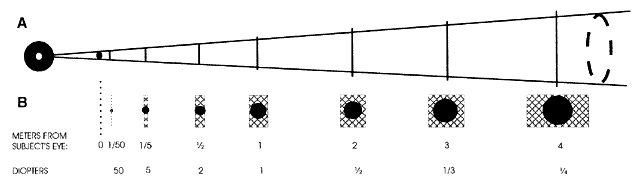
| Fig. 23. The relative magnifications and distances (from the subject's eye) of images of the retinoscope filament when observing the image “of the retina” in hyperopic eyes. The eye may be considered the hyperopic version of the schematic eye shown in Figure 16. The magnification is calculated as the ratio of distance from the image of the retina to the subject's pupil divided by distance from the pupil to the retina. A. Cone to which the observer's view is restricted by the subject's pupil. Its apex is at the observer's pupil (x) and its angular dimension is determined by the subject's pupil (y). The lengths of the vertical solid bars traversing the cone represent the widths of the images of the filament at several distances from the eye. The segment of the bar that lies within the cone is visible to the observer. B. Proportion of the filament image that can be seen by the observer represented in another form. The filament image (cross-hatched area) rapidly increases as the ametropia decreases. Beyond approximately 1.00 D, the edges of the filament image cannot lie within the cone, and enhancement cannot occur. Enhancement can occur in eyes with 2.00 D or more of hyperopia. The filament image seen through the subject's pupil is shown as a dark band. The retina is located 0.02 m from the subject's pupil. Consider the extreme example of + 50.00 D, in which the “image of the retina” is “on the retina.” It would appear as a very small and bright line that would not fill the pupil. A 50.00-D refraction at 1/50 of a meter = 2 cm. (Modified from Safir A: Retinoscopy. In Tasman W, Jaeger EA [eds]: Duane's Clinical Ophthalmology. Philadelphia: JB Lippincott, 1982.) |