|
|
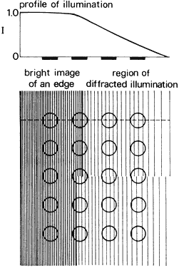
| Fig. 51. Vernier acuity. Diffracted energy on the retina from the edge of a broken line spreads out as shown by the profile of illumination, which corresponds to a scan along the dashed line. The profile for the upper region is slightly offset from that of the lower region; consequently, the cones in the columns above the break in the line receive more illumination than the cones below the break. |