|
|
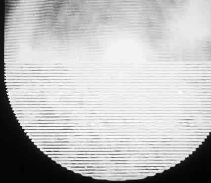
| Fig. 12. Interference fringes as they appear normally (top) and with double images focused on tissue paper placed over camera lens (bottom). Cataract patients may see continuously changing fringe patterns similar to those patterns on the bottom. (Green DG: Testing the vision of cataract patients by means of laser generated interference fringes. Science 16(June):5, 1970 [cover photo]. Copyright © 1970, American Association for the Advancement of Science) |