|
|
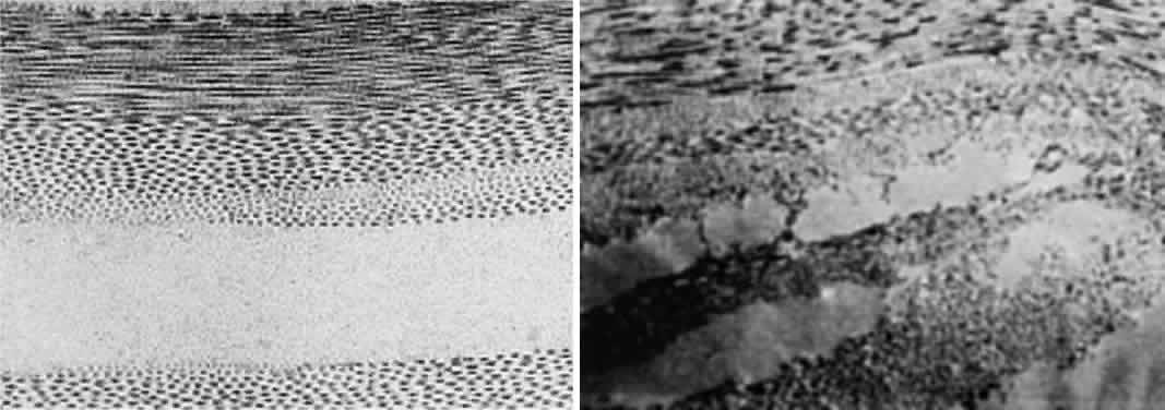
| Fig. 5. Top. Electron micrograph shows the arrangement of collagen fibers in normal corneal stroma. Bottom. Electron micrograph of a corneal stroma with edema. Note the irregular collection of fluid. (Miller D, Benedek G: Intraocular Light Scattering. Springfield, IL, Charles C Thomas, 1973. Courtesy of T. Kuwabara, Howe Laboratory, Harvard Medical School) |