|
|
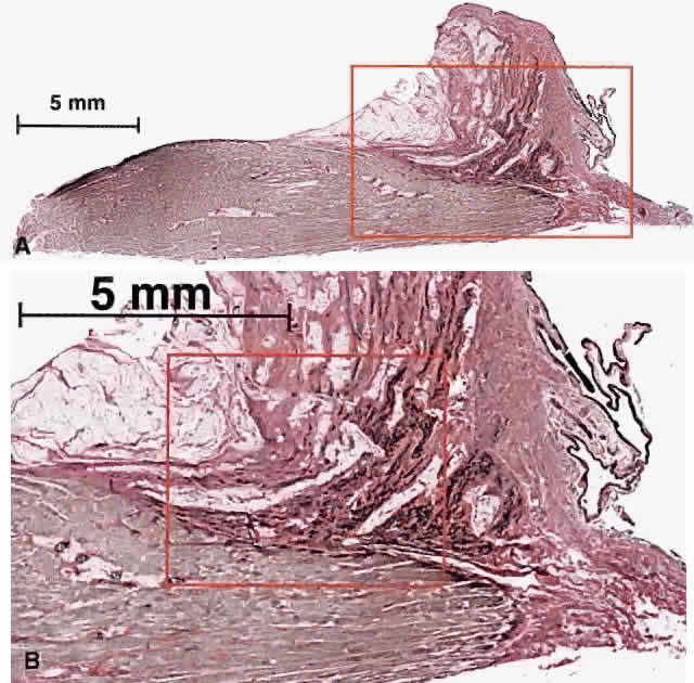
| Fig. 12. Longitudinal sections of human medial rectus (MR) pulley demonstrating insertion of the orbital layer on the pulley. A. Low-power micrograph of longitudinal section of human MR in continuity with pulley stained by van Giesson's method to demonstrate elastin fibers in dark black. Dense fascicles of elastin, embedded in collagen, run anteriorly from the pulley toward the anterior lacrimal crest. B. Moderate-power micrograph of longitudinal section of human MR in continuity with pulley in the region designated by the red rectangle in A, stained by van Giesson's method. Discrete elastin fibrils are evident. C. High-power micrograph of longitudinal section of human MR in continuity with pulley in the region designated by the red rectangle in B, stained by Masson's trichrome method. Dense collagen of the pulley is stained blue. Fascicles of smooth muscle in pulley suspension are indicated. (Micrograph by Dr. Sei Yeul Oh, MD, and the author.) |