|
|
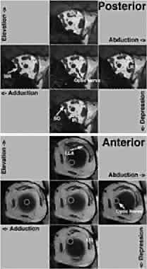
| Fig. 8. Coronal MRI of left orbit in primary position and in the secondary positions of abduction, adduction, elevation, and depression. IR, inferior rectus; LG, orbital lobe of lacrimal gland; LLA, lateral levator aponeurosis; LR, lateral rectus; MR, medial rectus; SR, superior rectus; SO, superior oblique. The upper five images were obtained posteriorly in the orbit, where increases in the cross sections of contracting rectus and SO extraocular muscles (EOMs; labeled) are most evident. At this level, EOM positions do not shift with gaze. The lower five images were obtained anteriorly in the orbit, at the level of the junction of the globe with the optic nerve just posterior to the pulleys, where rectus EOM paths do not shift appreciably with gaze and there is little contractile change in EOM cross sections. The shift in position of the optic nerve (white circle) is the only evidence of the large gaze shift. |