|
|
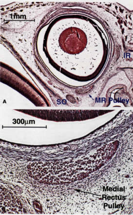
| Fig. 1. Coronal section of fetal human orbits stained with Masson's trichrome. A. Lower-power view of specimen at 11 weeks of gestation showing primary myogenesis of the extraocular muscles. Although no collagen is yet present, the medial rectus (MR) is encircled with connective tissue precursors that will form its pulley. Note the presence of both primary and secondary lens fibers in the lens. B. Higher-power view of 11-week specimen shows detail of pulley and MR muscle. IR, inferior rectus; SO, superior oblique. |