|
Please use the links below to immediately access specific areas of the protocol |
|
| Aims
To devise a system that is immediate, universally applicable and capable of accurately describing the range of blebs found after Trabeculectomy
Protocol:
Imaging the blebs.
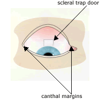
The system relies on the ability to provide standard photographs of the superior conjunctiva. It is hoped that most digital cameras can provide these photographs. The standard photograph will be a mono image with the eye looking inferiorly to display the largest area available of the superior conjunctiva. The canthal margins should be the horizontal limits of the photograph.
A standard photograph is displayed. |
| |
We recommend that the images be graded under magnification. Image magnification to a diagonal length of 15 inches, such as digitally enlarged images on a screen, would seem appropriate but individual centres are encouraged to use whatever imaging systems they have available.
|
|
Principles:
In this grading system the grader is asked to make judgements about the appearance of the bleb. Standard reference photos are provided. Where the bleb being graded does not match the reference photographs then the best match should be selected. Describing a bleb, three main aspects have to be considered: area, height and vascularity. There are six criteria to assess: 2 describing area, 1 describing height and 3 describing vascularity. |
 |
1) Area
The peripheral margins (maximal area) of the bleb, as well as the central demarcated area of the bleb are assessed.
1a: The central demarcated area of the bleb is compared with that of the total conjunctival area visible in the picture. Usually this area is located over the scleral trap door. On occasions when the entire bleb is uniform and not demarcated then the central area will equal the peripheral margins. |
| |
The area is given a score of 1-5 depending on its extension
1= 0% (even a small demarcated area, if the value is closest to 0%, as shown.)
2= 25%
3= 50%
4= 75%
5= 100%
The number given relates to the area that best describes what is found in the bleb |
|
2a: The whole, maximal area of the bleb (ie, its peripheral margins) is compared with that of the total area visible in the photograph of conjunctiva. The area is given a score of 1-5 depending on its extension.
1= 0%
2= 25%
3= 50%
4= 75%
5= 100%
The number given relates to the area that best describes what is found in the picture. The area of diffusion can be difficult to detect. The best visual clues are found under magnification and are elevation of the conjunctiva at the limbus forming a shallow limbal gutter. A "boggy" appearance caused by the elevation of the conjunctiva allows the superficial conjunctival vessels to be distinct and the deeper episcleral vessels are less well defined.
Reference photographs are given. |
|
2)Height
The height is compared to the height of the reference photographs provided and the value given which best fits the height of the bleb being graded. This is applied to the highest point of the bleb and usually represents the centre. Give a score between 1-4.
3)Vascularity
The vascularity relates to various parts of the bleb:
3a: the central demarcated area of the bleb; is the part of the bleb described in point 1a.
3b: the surrounding, peripheral part of the bleb (as described in 1b). When the bleb does not have a peripheral part (eg an encysted bleb with no surrounding diffusion) then the margin between central bleb and non-bleb should be described.
3c: the peripheral, non-bleb conjunctiva. When considering a bleb of large area (100%) then the peripheral non-bleb conjunctiva is not visible and the same value for 3b should be added.
Each part is compared to standard photographs provided. The number given to each part relates to the picture that best fits the height of the bleb being graded. The vascularity may vary over the bleb area and the value chosen should best represent the majority of the area being considered.
1: avascular
2: normal vascularization
3: mild vessel inflammation
4: moderate vessel inflammation
5: severe vessel inflammation
Where there is subconjunctival blood, this is to be graded with a note:
YES: if the subconjunctival blood is bigger than the scleral trap door
NO: if the subconjunctival blood is smaller than the scleral trap door (not clinically significant) or not found.
Reference photographs are provided. |
|
Glossary
Encysted bleb is one that has been walled off by the tenons capsule and therefore is elevated and tense in appearance.
Avascular blebs have areas of conjunctival thinning. The thin conjunctiva has no blood vessels. |
[1] [2] 下一页 |


 网友评论:(只显示最新10条。评论内容只代表网友观点,与本站立场无关!)